At NICT, we have
developed numerical human-body models with the aim of
evaluating the safety of radio waves with respect to the human
body, and are making them available to the public. At present, we
provide the voxel human model databases shown below.
Improvements
are also being made to these models, such as by adding specific
tissues and standardizing tissue weight, for example, and we plan
to successively update the database’s version. All version
updates will be announced on our website, and database users will
be contacted by email and other means.
Method of application for use
Research institutes such as universities
- Confirm the content of the draft of the database license contract.
- Fill in the necessary items in the Usage Application Form.
- We will contact you about your eligibility for use. If you are judged to be eligible, you will be asked to go through the database license contract procedures with Intellectual Property Management Group at NICT’s Research Promotion Department.
- After a contract has been concluded, we will send you encrypted data and a decoding password.
- If the purpose of your data use looks like it will not be accomplished by the time the database usage period expires, you may submit a request for extension of database usage before your usage period expires.
- If the purpose of your data use has been accomplished, or if the usage period has expired, submit a certificate of database deletion without delay.
Voxel human model database: Usage Application
-
Please fill in the necessary information below and apply by email. We will email you at a later date to notify you of your eligibility to use the database.
- [name]
- [title]
- [affilication]
- [address]
- [telephone]
- [email]
- [purpose_of_use]
- [period]
- [model or software]
(Adult male and female voxel models, Pregnant woman voxel model, Child models, Voxel Human Anatomy Lab, Please indicate the model and software you would like to use.)
E-mail: vhm(at)ml.nict.go.jp *Please replace (at) with @.
Private-sector corporations (those requesting usage for a fee)
At NICT, we have been distributing, free of charge, the voxel human model
database to research institutions such as
universities in both Japan and overseas, on condition that they be
used for non-commercial or research purposes only. The findings of
research using the database released at academic meetings, etc.,
have attracted the interest of numerous corporations and other
entities which until now were not eligible for database offerings.
At present, NICT distributes data to corporations and other
commercial entities as well, in the hope of having these data
utilized even more effectively.
For details on how to apply,
the license format and other information, contact Intellectual
Property Management Group.
Contact information for the department in charge
Intellectual Property Promotion Office, Innovation Promotion Department, NICT Tel: +81 42-327-5716 / Fax: +81 42-327-6659 / E-mail:ippo@ml.nict.go.jp
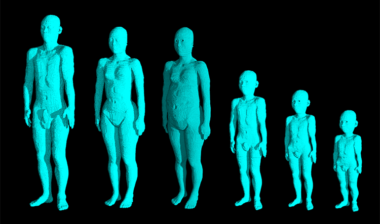
Adult male and female voxel models (Taro and Hanako)
Spatial resolution: 2 x 2 x 2 mm
No. of voxels: Male model:
approximately 8 million; female model: approximately 6.3 million
Comparison of average physique and model physique
| Average | Model | Margin of error (%) | ||
| Male | Height (cm) | 171.4 | 172.8 | +0.82 |
| Weight (kg) | 63.3 | 65.0 | +2.69 | |
| Female | Height (cm) | 159.1 | 160.0 | +0.57 |
| Weight (kg) | 52.6 | 53.0 | +0.76 | |
Average physique: The average values of subjects aged 18 to 30 featured in “The Japanese Body Dimensional Data for Ergonomic Design, 1996,” edited by the National Institute of Bioscience and Human Technology, and issued by Japan Publication Service.
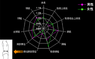
The physique of numerical human-body models
The numerical models were compared with the average physique of the Japanese people. Although both men and women showed certain deviations in the diameter of the naval-area abdominal thickness because of differences in posture during measurement of physique (numerical model: supine position; average value for Japanese people: standing position), the results show that the average difference between the numerical model and Japanese people’s average physique was below 5%, indicating that these numerical models coincide more or less with the average physique of Japanese people. (The closer to 1.0 the numerical figures in the diagram are, the more similar the two models are.)
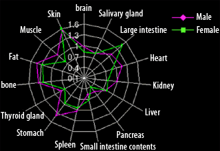
Weight of the tissues and organs of numerical human-body models
Although the organs showed certain individual differences, 60% of the tissues and organs of men and 80% of those of women differed from the average values by within 30%.
List of identified tissues and organs
| Name of tissue | |||
| Cerebellum | Esophagus | Uterus*1 | Cerebrospinal fluid |
| Bile | Air (in vivo space) | Cornea | Gall bladder |
| Blood | Eyeball | Heart | Cortical bone |
| Gray matter | Kidney | Bone marrow, cancellous bone | Hypothalamus |
| Liver | Cartilage | Crystalline lens | Lung |
| Fat | Epiphysis | Ovary*1 | Muscle |
| Pituitary gland | Pancreas | Nerve (spinal cord) | Salivary gland |
| Prostate gland*2 | Skin | Thalamus | Small intestine |
| Teeth | Tongue | Spleen | Ligament |
| White matter | Stomach | Small intestine contents | Adrenal gland |
| Stomach contents | Diaphragm | Bladder | Tendon |
| Seminal vesicle*2 | Mammary fat*1 | Testicle*2 | Cavernous body*2 |
| Large intestine | Thyroid gland | Vagina*1 | Large intestine contents |
| Trachea | Duodenum | Inside the bladder and urine | |
*1: Tissue unique to women 2: Tissue unique to men
Female pregnancy model
The female pregnancy model was developed by combining the fetus
model, which was produced from an abdominal MRI image of a woman
in the 26th week of gestation, with a model based on NICT’s
adult female model (Hanako) but expanding the abdominal area
three-dimensionally to match the body shape of a pregnant
woman.
The anatomical validity of this model was confirmed by
a specialist physician who provided medical supervision. We have
also confirmed that the dimensions of various parts of a pregnant
woman’s abdominal area, as well as the tissue and organ
weight specific to pregnant women, which include those of the
fetus, are close to the average values of women in their 26th week
of pregnancy.
Spatial resolution: 2 x 2 x 2 mm
Number of voxels:
Approximately 7.1 million
Fetus: 26th week of gestation
List of identified tissues and organs
| Names of tissues and organs | |||
| Cerebellum | Esophagus | Cortical bone | Cerebrospinal fluid |
| Bile | Bone marrow, cancellous bone | Cornea | Gallbladder |
| Cartilage | Eyeball | Heart | Fat |
| Gray matter | Kidney | Muscle | Hypothalamus |
| Liver | Nerves (spinal cord) | Crystalline lens | Lung |
| Skin | Epiphysis | Ovary*1 | Teeth |
| Pituitary gland | Pancreas | Tendon | Salivary gland |
| Small intestine | Small intestine contents | Thalamus | Spleen |
| Diaphragm | Tongue | Stomach | Vagina*1 |
| White matter | Stomach contents | Uterine wall*1 | Adrenal gland |
| Tendon | Fetus*2 | Bladder | Thyroid gland |
| Fetus’ brain*2 | Mammary fat*1 | Trachea | Fetus’ eyeballs*2 |
| Large intestine | Inside the bladder and urine | Amniotic fluid*2 | Large intestine contents |
| Air (in vivo space) | Placenta*2 | Duodenum | Blood |